Carotid Artery Disease
Normal blood flow to the brain is supplied by the carotid arteries. Like
other arteries in the vascular system, the carotid arteries can become
diseased with cholesterol plaque, a process called atherosclerosis. Cholesterol
deposits accumulate in the inner lining of the carotid artery and cause
gradual narrowing of the artery channel. This process is progressive and
occurs over a period of years.
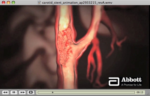
As the carotid plaque becomes more severe, a clot may form at its surface,
then break off and travel to the small arterioles of the brain, plugging
them and blocking blood flow to a portion of the brain. This causes oxygen
deprivation (also known as ischemia) to the brain cells, and may cause
the sudden onset of neurological symptoms. Some of these symptoms include
weakness or loss of strength of the arm or leg, or of the facial muscles,
numbness, difficulty speaking, or loss of vision in a portion of the visual
field. If the clot is rapidly dissolved by the body’s built-in clot
removal system, the oxygen deficiency is transient, the symptoms remit,
and no permanent damage occurs to the brain (thus, a CT scan or MRI will
not show any abnormality). This is called transient ischemic attack (TIA).
If the occlusion lasts more than a few hours, however, permanent damage
occurs to a portion of the brain. This is a stroke, or cerebrovascular
accident (CVA). A CVA causes changes in the brain tissue that can be detected
by a CT scan or MRI.
Diagnosing Carotid Artery Disease
 Carotid artery disease can be suspected during a physical exam, when a
physician listens with a stethoscope over your carotid artery, on each
side of the neck. When a blockage is present in the carotid artery, the
turbulence caused by interference to the blood flow causes a sound (“bruit”)
that can be heard with a stethoscope.
Carotid artery disease can be suspected during a physical exam, when a
physician listens with a stethoscope over your carotid artery, on each
side of the neck. When a blockage is present in the carotid artery, the
turbulence caused by interference to the blood flow causes a sound (“bruit”)
that can be heard with a stethoscope.
Carotid artery disease can be diagnosed by several kinds of tests. The
principal test used for this purpose is the carotid artery ultrasound.
This test is performed in a vascular ultrasound lab, is painless, and
takes about 30 min. In this test, the carotid artery, and blood flow through
the carotid, are examined with ultrasound imaging. A carotid ultrasound
can provide a very precise assessment of the health of the carotid artery.
An imaging test that is sometimes valuable as a screening tool is an examination
of the Intima-Media Thickness (IMT) of the carotid artery. When atherosclerosis
develops, the earliest events are the appearance of microscopic deposits
of cholesterol plaque in the inner lining of the carotid. This causes
the inner lining (the intima) to begin to thicken. In this test, the carotid
artery is examined with ultrasound, and the thickness of the intima layer
is carefully measured. Thus, this test detects the atherosclerosis process
in its early stages, before it is fully manifest as a significant blockage.
This data can be helpful information to you, as you can detect atherosclerosis
early and make changes to modify your future risk. The test costs about
$250. Unfortunately, this test is not covered by Medicare and many insurance
companies.
Another imaging modality is CT angiography or MR angiography, where the
carotid artery is examined by CT or MRI scanning. This test uses IV iodine
contrast (CT) or gadolinium contrast (MRI) to image the carotid artery.
The test requires an IV line. Some patients with kidney problems may not
be able to tolerate iodine or gadolinium contrast.
Lastly, direct imaging of the carotid arteries can be done with formal
angiography. During an angiogram, a catheter is inserted in the groin
and advanced into the carotid artery. IV iodine contrast is then injected
under x-ray visualization, and the artery is imaged.
Treating Carotid Artery Disease
The treatment strategy for carotid artery disease is based on the severity
of the blockage, and whether or not you have ever had any symptoms, such
as TIA or stroke:
The more severe the blockage, the more likely you will need to have some
kind of invasive therapy.
A blockage that has already caused some clinical event, such as a TIA or
stroke, will need more aggressive treatment than one that has not caused symptoms.
Mild blockages <60% can be treated with medications. These include antiplatelet
agents (such as aspirin, clopidogrel, prasugrel, dipyridamole) and cholesterol-lowering
drugs (mainly in the statin family — lovastatin, simvastatin, pravastatin,
atorvostatin, etc.).
More severe blockages >60% generally are treated with invasive therapy:
carotid endarterectomy or carotid stenting. In carotid endarterectomy
(CEA), typically done under general anesthesia, a vascular surgeon makes
an incision in the neck, then opens the carotid artery and removes the
plaque from the carotid artery, then closes the artery with suture. Typically,
this requires a 2 or 3 day hospitalization.
A carotid stent procedure, though considered invasive, is less invasive
than CEA. This procedure, similar to a carotid angiogram, is performed
through a needle hole in the femoral artery at the groin. Typically, the
carotid stent procedure is performed immediately following a carotid angiogram;
i.e., in the same setting. A catheter is inserted through the groin and
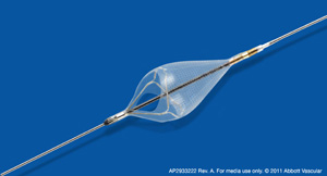
its tip is positioned at the origin of the carotid artery. A small filter
basket is then inserted into the distal portion of the carotid artery,
beyond the blockage, to “catch” any debris that may be generated
during the procedure. A balloon is then positioned at the site of the
blockage and briefly inflated to stretch open the blockage. A stent, made
of nickel-titanium, is then placed at the blockage to keep the artery
open. The procedure takes about 1-2 hours. A carotid stent procedure typically
requires an overnight hospitalization.
The decision on medical therapy versus invasive therapy, and which specific
type of invasive therapy, depends on unique characteristics. These include
carotid anatomy, presence or absence of other conditions, such as cardiac,
pulmonary, or renal disease. Every patient is different, and the best
therapy for you is something that you and your doctor should tailor to
your particular situation and needs. A detailed discussion with your doctor
is central to this decision making process.